2.2 Strong-field Few-body Physics

Project coordinators: J. Mikosch, F. Morales Moreno, B. Schuette

Recent Highlights

Photon-recoil imaging: a new route to study nonlinear x-ray physics

One key scientific driver for the development of intense x-ray free-electron lasers (XFELs) has been the prospect to transfer the concepts of nonlinear optics into the x-ray spectral range. Now scientists from the Max Born Institute (MBI), Uppsala University and the European XFEL have expanded the view on how to look at nonlinear x-ray processes. In a study published in Science, they investigated stimulated x-ray Raman scattering in a truly innovative way. Rather than detecting the scattered x-ray radiation from dense (gaseous) media as it is usually pursued in related studies they measured individual scattered atoms subjected to the photon recoil.

In one of the first user experiments at the SQS instrument at the European XFEL in Hamburg the German-Swedish collaboration has established the photon-recoil imaging technique to distinguish spontaneous from nonlinear stimulated x-ray Raman scattering. To understand the new technique it is helpful to recall that a photon, i.e., the quantized energy portion of electromagnetic radiation, also carries momentum; a fact that is well-known since the early days of quantum physics at the times of Einstein.  As a consequence, absorption of a photon inevitably pushes the atom very much like a billiard ball gets a push when hit by another one. In the spontaneous Raman scattering process, transient absorption of a photon is rapidly followed by spontaneous emission of a photon with less energy. The photons’ energy difference remains in the atom lifting an electron to a bound excited state. While the absorption pushes the atom in the direction of the incoming photon, the spontaneous emission of a photon, which happens into a random direction, scatters the atom accordingly to a corresponding opposite random direction.

To detect the scattered excited atoms in the experiment the scientists used a collimated supersonic beam of neon atoms, which travel towards a position-sensitive detector. The detector is set to be sensitive to impinging excited atoms only. Well in front of the detector the atomic beam is crossed perpendicularly by the XFEL beam defining a sharp elongated interaction volume. If the x-ray photon energy is tuned close to an inner-shell resonance of the Ne atom, about 2 % of the transiently excited atoms undergo spontaneous x-ray Raman scattering leaving the atom intact in finally a long lived bound excited state (the overwhelming portion of transiently excited atoms ionizes through a fast Auger process since the transient excitation energy exceeds the first ionization threshold by a factor of about 40 ). The photon momentum transfer slightly deflects the excited atoms from the collimated atomic beam. Accumulating the resulting detector signal over many XFEL pulses a characteristic pattern on the detector is produced, as shown in Fig. 1a. The pattern has an extended elliptical shape due to the deflections in random directions experienced by the atoms in the interaction volume during spontaneous Raman scattering.
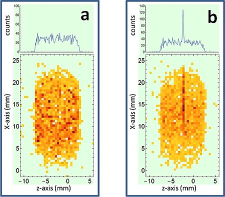
Figure 1:

a) Distribution of excited atoms on the detector after spontaneous x-ray Raman scattering

b) Same as a) plus excited atoms after stimulated x-ray Raman scattering (narrow stripe)

When the scientists optimized the XFEL intensity and the photon energy of the x-ray beam, which has a photon energy bandwidth in the range of the atomic excitation energy, they increased the probability for the transiently excited atom to get hit by a second photon with suitable energy, before it actually has a chance to emit a photon spontaneously. In this case the transiently excited atom will be stimulated to emit a photon along the direction of the incoming photon. Since this process depends on two photons of the x-ray pulse, the process is called nonlinear. The net momentum transfer by the incoming and stimulated photon is almost zero due to the low final excitation energy of the atom compared with the x-ray photon energies. Excited atoms from stimulated Raman scattering are thus basically not deflected and appear on the detector as a sharp straight line mapping the interaction volume. They can be clearly distinguished from the spontaneously Raman scattered atoms, as shown in Fig. 1b. The measured excited Ne* atom yield is amenable to a quantitative theoretical analysis as presented in the study.

Photon-recoil imaging enables the investigation of x-ray stimulated Raman scattering on a single atom level almost background free. In combination with short, time-delayed controlled pairs of x-ray pulses with different photon energies - which have already been demonstrated at seeded XFELs - it establishes a highly promising nonlinear x-ray spectroscopy method. The scientists envisage photon-recoil imaging to unravel fundamental features in x-ray photon interaction with atoms and molecules, even allowing to adopt coherent optical control methods.

Original publication

Photon-Recoil Imaging: expanding the view of nonlinear X-ray physics

U. Eichmann, H. Rottke, S. Meise, J.-E. Rubensson, J. Söderström, M. Agåker, C. Såthe, M. Meyer, T. M. Baumann, R. Boll, A. D. Fanis, P. Grychtol, M. Ilchen, T. Mazza, J. Montano, V. Music, Y. Ovcharenko, D. E. Rivas, S. Serkez, R. Wagner, and S. Eisebitt

Science Vol. 369, Issue 6511 (2020) pp. 1630-1633

URL, DOI or PDF

Overview MBI-Projects Overview Topics