Ultra-strong, ultra-fast and local: water induces electrical fields at the DNA surface

The structure and dynamics of the DNA double helix are influenced in a decisive way by the surrounding water shell. New experiments in the ultrafast time domain show that the first two water layers at the DNA surface generate electric fields of up to 100 megavolts/cm which fluctuate on the femtosecond time scale and are limited to a spatial range on the order of 1 nm.

DNA molecules are the carrier of genetic information and form a double helix structure in their native aqueous environment. This structure consists of two opposite twisted strands of nucleotides (Fig. 1A). An alternating arrangement of negatively charged phosphate groups and polar sugar units forms the backbone of the double helix structure which interacts directly with the surrounding water molecules. The overall negative charge of the double helix is compensated for by positively charged counterions such as sodium ions which in an aqueous environment are located in direct proximity to the DNA surface. The interaction of the electric dipoles of water molecules with the charges of the counterions and phosphate groups as well as the polar units generates electric fields at the DNA surface. Such fields are being discussed in a highly controversial way, even after decades of intense research, reflecting the structural complexity of this many-body system and its thermal fluctuations on short time scales.

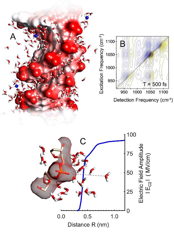
Fig. 1: (A) Surface of a DNA double helix. The twisted strands of the helix are represented by oxygen atoms (red) of the phosphate groups. Counterions are shown in blue, the small angled structures are water molecules. (B) Two-dimensional infrared spectrum of the DNA backbone. Nonlinear vibrational signals are plotted as a function of excitation and detection frequency. The lineshapes of the resonances on the diagonal (identical excitation and detection ferquency) are directly influenced by fluctuating electric fields. The off-diagonal signals originate from couplings between different backbone vibrations. (C) Time-averaged electric field (blue) as a function of the distance from the DNA surface. Water molecules in the first layer (around 0.4 nm) generate approximately 70% of the total field, the second water layer contributes some 20%.

Scientists from the Max-Born-Institute in Berlin have now succeeded for the first time in determining the strength, range, and ultrafast dynamics of electric fields at a native DNA surface. In a recent paper published in the Journal of Physical Chemistry Letters, they report how vibrations of the backbone of native salmon DNA serve as probes for mapping electric interactions in space and time. Electric fields at the surface directly influence the shape and dynamics of vibrational resonances which are recorded in real-time on the femtosecond time scale (1 fs = 10-15) s by a method referred to as two-dimensional infrared spectroscopy (Fig. 1B). The water content of the DNA samples has been varied in a systematic way to discern different contributions to the fluctuating electric fields at the DNA surface. 

The experiments and a detailed theoretical analysis performed in parallel show that water molecules in the first two layers at the DNA surface generate an extremely strong electric field whereas the ionic groups and outer water layers play a minor role. The spatial range of the field is approximately 1 nm, its strength reaches values up to 100 megavolts/cm (hundred million volts per centimeter) as shown in Fig. 1C. Thermal motions of the water molecules result in field fluctuations of 25 MV/cm on a 300 fs time scale. The time scale of fluctuations demonstrates a hindrance of water motions due to the coupling to the corrugated DNA surface and a slowing down compared to neat water. This new quantitative insight is important for understanding the key role of water and its dynamics at biological interfaces such as charged cell membranes and the surface of proteins. 

Search publications of MBI

advanced search
Search results

Publications since 2025

Sort: Year Author Title Journal
A1-P-2025.01
Melting, bubblelike expansion, and explosion of superheated plasmonic nanoparticles

S. Dold, T. Reichenbach, A. Colombo, J. Jordan, I. Barke, P. Behrens, N. Bernhardt, J. Correa, S. Düsterer, B. Erk, T. Fennel, L. Hecht, A. Heilrath, R. Irsig, N. Iwe, P. Kolb, B. Kruse, B. Langbehn, B. Manschwetus, P. Marienhagen, F. Martinez, K.-H. Meiwes-Broer, K. Oldenburg, C. Passow, C. Peltz, M. Sauppe, F. Seel, R. M. P. Tanyag, R. Treusch, A. Ulmer, S. Walz, M. Moseler, T. Möller, D. Rupp, B. v. Issendorff

Physical review letters 134 (2025) 136101/1-7

URL, DOI or PDF

A3-P-2025.01
Second-harmonic generation in OP-GaAs0.75P0.25 heteroepitaxially grown from the vapor phase

L. Wang, S. R. Vangala, S. Popien, M. Beutler, J. M. Mann, V. L. Tassev, E. Büttner, V. Petrov

CrystEngComm 27 (2025) 1373-1376

URL, DOI or PDF

A3-P-2025.02
Diode-pumped Kerr-lens mode-locked Yb:MgWO4 laser

H.-Y. Nie, Z.-L. Lin, P. Loiko, H.-J. Zeng, L. Zhang, Z. Lin, G. Z. Elabedine, X. Mateos, V. Petrov, G. Zhang, W. Chen

Optics Letters 50 (2025) 1049-1052

URL, DOI or PDF

A3-P-2025.03
Growth, anisotropy, and spectroscopy of Tm3+ and Yb3+ doped MgWO4 crystals

G. Z. Elabedine, R. M. Solé, S. Slimi, M. Aguiló, F. Díaz, W. Chen, V. Petrov, X. Mateos

CrystEngComm 27 (2025) 1619-1631

URL, DOI or PDF

A3-P-2025.04
Growth, structure, spectroscopic, and laser properties of Ho-doped yttrium gallium garnet crystal

S. Slimi, H. Yu, H. Zhang, C. Kränkel, P. Loiko, R. M. Solé, M. Aguiló, F. Díaz, W. Chen, U. Griebner, V. Petrov, X. Mateos

Optics Express 33 (2025) 2529-2541

URL, DOI or PDF

A3-P-2025.05
Growth, spectroscopy and laser operation of disordered Tm,Ho:NaGd (MoO4)2 crystal

G. Z. Elabedine, Z. Pan, P. Loiko, H. Chu, D. Li, K. Eremeev, K. Subbotin, S. Pavlov, P. Camy, A. Braud, S. Slimi, R. M. Solé, M. Aguiló, F. Díaz, W. Chen, U. Griebner, V. Petrov, X. Mateos

Journal of Alloys and Compounds 1020 (2025) 179211/1-12

URL, DOI or PDF

A3-P-2025.06
Kerr-lens mode-locked, diode-pumped Yb,Gd:YAP laser generating 23 fs pulses

H.-Y. Nie, P. Zhang, P. Loiko, Z.-L. Lin, H.-J. Zeng, G. Zhang, Z. Li, X. Mateos, H.-C. Liang, V. Petrov, Z. Chen, W. Chen

Optics Express 33 (2025) 11793-11799

URL, DOI or PDF

A3-P-2025.07
Nanoindentation and laser-induced optical damage tests of CdSe nonlinear crystals

G. Exner, A. Carpenter, K. Cissner, A. Hildenbrand-Dhollande, S. Schmitt, A. Grigorov, M. Piotrowski, S. Guha, V. Petrov

Journal of the Optical Society of America B 42 (2025) A10-A14

URL, DOI or PDF

A3-P-2025.08
Phase-matching properties of AgGa(Se1-xTex)2 for SHG of a CO2 laser

K. Kato, V. Petrov, K. Miyata

Proceedings of SPIE 13347 (2025) 133470S/1-4

URL, DOI or PDF

A3-P-2025.09
Phase-matching properties of ZnSiAs2 in the mid-IR

T. Okamoto, N. Umemura, K. Kato, V. Petrov

Proceedings of SPIE 13347 (2025) 133470C/1-5

URL, DOI or PDF

A3-P-2025.10
Direct generation of 3.5 optical-cycle pulses from a rare-earth laser

N. Zhang, Y. Wang, H. Ding, F. Liang, Y. Zhao, J. Xu, H. Yu, H. Zhang, V. Petrov

Optics Letters 50 (2025) 3150-3153

URL, DOI or PDF

A3-P-2025.11
Power scaling of a non-resonant optical parametric oscillator based on periodically poled LiNbO3 with spectral narrowing

S. Das, T. Temel, G. Spindler, A. Schirrmacher, I. B. Divliansky, R. T. Murray, M. Piotrowski, L. Wang, W. Chen, O. Mhibik, V. Petrov

Optics Express 33 (2025) 5662-5669

URL, DOI or PDF

A3-P-2025.12
Sub-40-fs diode-pumped ytterbium-doped mixed rare-earth calcium oxoborate laser

H.-J. Zeng, Z.-L. Lin, H. Lin, P. Loiko, L. Zhang, Z. Lin, H.-C. Liang, X. Mateos, V. Petrov, G. Zhang, W. Chen

Optics Express 33 (2025) 17965-17975

URL, DOI or PDF

A3-P-2025.13
Spectroscopy and SESAM mode-locking of a disordered Yb:Gd2SrAl2O7 crystal

H.-J. Zeng, Z.-L. Lin, P. Loiko, F. Yuan, G. Zhang, Z. Lin, X. Mateos, V. Petrov, W. Chen

Optics Express 33 (2025) 15057-15066

URL, DOI or PDF

A3-P-2025.14
Watt-level, 1.6 ps χ(2)-lens mode-locking of an in-band pumped Nd:LuVO4 laser

H. Iliev, V. Aleksandrov, V. Petrov, L. S. Petrov, H. Zhang, H. Yu, I. Buchvarov

Optics Express 33 (2025) 17773-17781

URL, DOI or PDF

A3-P-2025.15
Refined phase-matching predictions for AgGa1-xInxS2 mixed chalcopyrite crystals

K. Kato, K. Miyata, V. Petrov

Journal of the Optical Society of America B 42 (2025) A6-A9

URL, DOI or PDF

A3-P-2025.16
35-fs diode-pumped mode-locked ytterbium-doped multi-component alkaline-earth fluoride laser

Z. Zhang, Z.-Q. Li, P. Loiko, H.-J. Zeng, G. Zhang, Z.-L. Lin, S. Normani, A. Braud, F. Ma, X. Mateos, H.-C. Liang, V. Petrov, D. Jiang, L. Su, W. Chen

Optics Letters 50 (2025) 1835-1838

URL, DOI or PDF

A3-P-2025.17
Diode-pumped few-optical-cycle laser based on an ytterbium-doped disordered strontium yttrium borate crystal

H. Zeng, Z. Lin, S. Sun, P. Loiko, H. Lin, G. Zhang, Z. Lin, C. Mou, X. Mateos, V. Petrov, W. Chen

Optics Letters 50 (2025) 2203-2206

URL, DOI or PDF

A3-P-2025.18
Refined Sellmeier and thermo-optic dispersion formulas for CdGeAs2

K. Kato, K. Miyata, V. Petrov

Journal of the Optical Society of America B 42 (2025) A24-A28

URL, DOI or PDF

A3-P-2025.19
Diode-pumped mode-locked Yb:Ca3La2(BO3)4 laser generating 35 fs pulses

H.-J. Zeng, Z.-L. Lin, G. Zhang, Z. Pan, P. Loiko, X. Mateos, V. Petrov, H. Lin, W. Chen

Optics Express 33 (2025) 22988-22996

URL, DOI or PDF